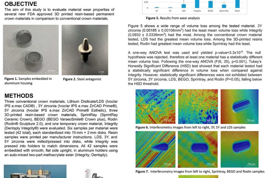
Objectives: Tooth movement is caused by the application of force. In short, applied force strains structures present in the PDL space – cells, ligaments, blood vessels. Cells in the PDL are damaged by extension and by diminished oxygen supply due to compression of blood vessels. Compounds released from damaged or dead cells trigger an innate inflammatory response. One of the biomarkers of that response is increased formation of extracellular fluid (edema), specifically gingival crevicular fluid (GCF). Our goal is to monitor changes of GCF flow during the orthodontic treatment with Invisalign.

Objectives: The purpose of this study is to compare the shear-bond strength of a chemically-cured bulk composite and a light-cured bulk composite. The chemically-cured bulk composite was delivered at room temperature. The light-cured composite was delivered at room temperature and at 155 degrees Fahrenheit. The null hypothesis is that there is no difference among the three groups.

Methods: 10 freshly extracted third molars were cross sectioned and embedded in resin to fabricate a specimen with a dimension of 2cm in height and 2.5cm in diameter. Group A: 10 specimens were used for shear bond strength measurement with the chemically-cured composite Bulk Easy +(Danville). Group B: 10 specimens were tested with light-cured composite Filtek Bulk One(3M) at room temperature. Group C: 10 specimens were tested with the light-cured composite Filtek Bulk One(3M) at 155-degrees Fahrenheit. The same 10 samples were reused for all groups. Shear bond testing was done with the Unitester(Ultradent) at a crosshead speed of 1 mm/min. Paired Student T-tests were performed to compare Group A with Group B, Group A with Group C and Group B with Group C with a 95% confidence level.

Results: The average results from the shear bond testing were 15.97 MPa for the chemically-cured composite(Group A), 23.07 MPa for the light-cured composite at room temperature(Group B), and 21.76 MPa for the light-cured composite warmed to 155 degrees Fahrenheit(Group C). Paired student T-tests between each group were done with a 95% confidence level. The p-value between Group A and B was 0.008, between Group A and C was 0.037, and between Group B and C was 0.266.

Conclusions: The chemically-cured composite has significantly lower shear-bond strength than both light-cured composite groups. No statistical difference was found between the light-cured groups.

Acknowledgements: We thank 3M, Zest Dental, and Bioclear for the materials used

Dental adhesives are water soluble, non-toxic materials that bind composite resins to dentin and enamel. Adhesive systems are dependent on a polymerization process at the interface and require different moieties to prepare the substrate to effectively interact with the tooth surface. Due to their critical role in restorative procedures, many chemists are interested in strengthening the adhesive bond strength at the interface. The review paper I published in the Pacific Journal of Health covers the evolution of dental adhesive systems over time and provides an extensive overview of what components make up a dental adhesive system. Additionally, the article explores the common issues found in adhesive systems, and how current researchers are working to approach these complications to increase the success rate of dental restorations.

One of the biggest barriers to care in providing oral health care is the fear of dentists from the public. Patients with special healthcare needs may have sensory triggers, anatomical abnormalities, and face other barriers when seeking dental care. When dental needs are unmet, the periodontal condition worsen, and could lead to unwanted tooth decay. Early intervention and maintenance care is important to ensure the patient’s oral health is in good condition. Intervention such as sensory desensitization, caregiver education, removing the barriers to seek care, and utilization of alternative tools such as a universal soft tissue retractor will help increase patient compliance.

The sounds, sights, and smells of a dental office have always been at the forefront of patient dental anxiety. With this in mind, we set out to identify how deafening dental instruments can be. The dental handpieces, suction devices, and hygiene equipment such as the cavitron dental scaler noise levels were measured. In this study, both the hearing of the dentist and patient were analyzed along with the overall decibel level and frequencies to determine what is causing the most negative effect on the hearing of the patient as well as the operator and dental personnel. The materials used in this study were: saliva ejector, high speed evacuation suction, cavitron, slow speed handpiece, electric high speed handpiece, and air driven high speed handpiece. We used a combination of dental instruments simulating what is used in a clinical setting. The collaboration between dentists, dental students, audiologists, and audiology students was strategically used in this study to allow for this study to be highly effective and specialized.